Bacteria

Bacteria constitute a large domain of prokaryotic microorganisms. Typically a few micrometres in length, bacteria have a number of shapes, ranging from spheres to rods and spirals. Bacteria were among the first life forms to appear on Earth, and are present in most of its habitats. Bacteria inhabit soil, water, acidic hot springs, radioactive waste,[4] and the deep portions of Earth’s crust. Bacteria also live in symbiotic and parasitic relationships with plants and animals. They are also known to have flourished in manned spacecraft.[5]

There are typically 40 million bacterial cells in a gram of soil and a million bacterial cells in a millilitre of fresh water. There are approximately 5×1030 bacteria on Earth,[6] forming a biomass which exceeds that of all plants and animals.[7] Bacteria are vital in recycling nutrients, with many of the stages in nutrient cycles dependent on these organisms, such as the fixation of nitrogen from the atmosphere and putrefaction. In the biological communities surrounding hydrothermal vents and cold seeps, bacteria provide the nutrients needed to sustain life by converting dissolved compounds, such as hydrogen sulphide and methane, to energy. On 17 March 2013, researchers reported data that suggested bacterial life forms thrive in the Mariana Trench, which with a depth of up to 11 kilometres is the deepest part of the Earth’s oceans.[8][9] Other researchers reported related studies that microbes thrive inside rocks up to 580 metres below the sea floor under 2.6 kilometres of ocean off the coast of the northwestern United States.[8][10] According to one of the researchers, “You can find microbes everywhere — they’re extremely adaptable to conditions, and survive wherever they are.”[8]

Most bacteria have not been characterized, and only about half of the phyla of bacteria have species that can begrown in the laboratory.[11] The study of bacteria is known as bacteriology, a branch of microbiology.

There are approximately ten times as many bacterial cells in the human flora as there are human cells in the body, with the largest number of the human flora being in the gut flora, and a large number on the skin.[12] The vast majority of the bacteria in the body are rendered harmless by the protective effects of the immune system, and some are beneficial. However, several species of bacteria are pathogenic and cause infectious diseases, including cholera, syphilis, anthrax, leprosy, and bubonic plague. The most common fatal bacterial diseases arerespiratory infections, with tuberculosis alone killing about 2 million people per year, mostly in sub-Saharan Africa.[13] In developed countries, antibiotics are used to treat bacterial infections and are also used in farming, making antibiotic resistance a growing problem. In industry, bacteria are important in sewage treatment and the breakdown of oil spills, the production of cheese and yogurt through fermentation, and the recovery of gold, palladium, copper and other metals in the mining sector,[14] as well as in biotechnology, and the manufacture of antibiotics and other chemicals.[15]

Once regarded as plants constituting the class Schizomycetes, bacteria

are now classified as prokaryotes. Unlike cells of animals and other eukaryotes, bacterial cells do not contain a nucleus and rarely harbour membrane-bound organelles. Although the term bacteria traditionally included all prokaryotes, the scientific classificationchanged after the discovery in the 1990s that prokaryotes consist of two very different groups of organisms thatevolved from an ancient common ancestor. These evolutionary domains are called Bacteria and Archaea.[1]

Formation of Tornadoes

Step 1: Like all winds and storms, tornadoes begin when the sun heats up the surface of the land. As the warm, less heavy air begins to rise, it meets the colder, heavier air above it. Note that wind shears make it even easier to set them off. A wind shear is when two winds at different levels and speeds above the ground blow together in a location.

Step 2: The faster moving air begins to spin and roll over the slower wind. As it rolls on, it gathers pace and grow in size.

Step 3: At this stage, it is an invisible, horizontal wind spinning and rolling like a cylinder. As the winds continue to build up, stronger and more powerful warm air forces the spinning winds vertically upward, causing an updraft.

Step 4: With more warm air rising, the spinning air encounters more updraft. The winds spin faster, vertically upwards, and gains more momentum.

Step 5: At this stage, the spinning winds, creates a vortex and the the wind has enough energy to fuel itself.

Step 6: The tornado is fully formed now and moving in the direction of the thunderstorm winds. When the pointed part of the tornado touched the ground from the cloud, it is often referred to as ‘touch down’ As it moves it rips off things along its patch.

How to add whole numbers?

Addition is to add two or more numbers to get the total number.

Example 1:

Add 1+1

Here 1 ball is added

to 1 ball

to make 2 balls:  


Example 2: 

Add 2+3

Let say that you have 2 green boxes and you have 3 blue boxes and count all the boxes and the total number of boxes is 5 boxes.

2+3=5

Example 3:

  
As shown in the figure, there are 4 blue balls and 3 red balls. Just simply count all the balls in the figure to get the answer or add the blue balls and the red balls.

4 blue balls + 3 red balls = 7 balls

Example 4:

Add 6+9

 6+9=15

Example 5:

12+13

Add the ones place (2 and 3)

2+3=5

Add the tens place (both 1)

1+1=2

Combine the tens and ones place

12+13=25

2+3=5

Displacement

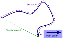
A displacement is the shortest distance from the initial to the final position of a point P.[1] Thus, it is the length of an imaginary straight path, typically distinct from the path actually travelled by P. A displacement vector represents the length and direction of this imaginary straight path.

A position vector expresses the position of a point P in space in terms of a displacement from an arbitrary reference point O (typically the origin of a coordinate system). Namely, it indicates both the distance and direction of an imaginary motion along a straight line from the reference position to the actual position of the point.

A displacement may be also described as a ‘relative position’: the final position of a point (Rf) relative to its initial position (Ri), and a displacement vector can be mathematically defined as the difference between the final and initial position vectors:


\boldsymbol{s}=\boldsymbol{R_f-R_i}=\Delta\boldsymbol{R}

In considering motions of objects over time the instantaneous velocity of the object is the rate of change of the displacement as a function of time. The velocity then is distinct from the instantaneous speed which is the time rate of change of the distance traveled along a specific path. The velocity may be equivalently defined as the time rate of change of the position vector. If one considers a moving initial position, or equivalently a moving origin (e.g. an initial position or origin which is fixed to a train wagon, which in turn moves with respect to its rail track), the velocity of P (e.g. a point representing the position of a passenger walking on the train) may be referred to as a relative velocity, as opposed to an absolute velocity, which is computed with respect to a point which is considered to be ‘fixed in space’ (such as, for instance, a point fixed on the floor of the train station).

For motion over a given interval of time, the displacement divided by the length of the time interval defines the average velocity. (Note that the average velocity, as a vector, differs from the average speed that is the ratio of the path length — a scalar — and the time interval.)

Displacement versus distance traveled along a path

Rigid body[edit]

In dealing with the motion of a rigid body, the term displacement may also include the rotations of the body. In this case, the displacement of a particle of the body is called linear displacement (displacement along a line), while the rotation of the body is called angular displacement.

Derivatives[edit]

For a position vector s that is a function of time t, the derivatives can be computed with respect to t. These derivatives have common utility in the study ofkinematics, control theory, vibration sensing and other sciences and engineering disciplines.

Velocity

\boldsymbol{v}=\frac{\text{d}\boldsymbol{s}}{\text{d}t} (where ds is an infinitesimally small displacement)

Acceleration

\boldsymbol{a}=\frac{\text{d}\boldsymbol{v}}{\text{d}t}=\frac{\text{d}^2\boldsymbol{s}}{\text{d}t^2}

Jerk

\boldsymbol{j}=\frac{\text{d}\boldsymbol{a}}{\text{d}t}=\frac{\text{d}^2\boldsymbol{v}}{\text{d}t^2}=\frac{\text{d}^3\boldsymbol{s}}{\text{d}t^3}

These common names correspond to terminology used in basic kinematics.[2] By extension, the higher order derivatives can be computed in a similar fashion. Study of these higher order derivatives can improve approximations of the original displacement function. Such higher-order terms are required in order to accurately represent the displacement function as a sum of an infinite series, enabling several analytical techniques in engineering and physics

Glucose

Glucose is a sugar with the molecular formula C6H12O6. The name “glucose” (/ˈɡlks/) comes from the Greek word γλευκος, meaning “sweet wine, must“.[3] The suffix “-ose” is a chemical classifier, denoting a carbohydrate. It is also known as dextrose or grape sugar. With 6 carbon atoms, it is classed as a hexose, a sub-category ofmonosaccharides. α-D-glucose is one of the 16 aldose stereoisomers. The D-isomer (D-glucose) occurs widely in nature, but the L-isomer (L-glucose) does not. Glucose is made during photosynthesis from water and carbon dioxide, using energy from sunlight. The reverse of the photosynthesis reaction, which releases this energy, is a very important source of power for cellular respiration. Glucose is stored as a polymer, in plants as starch and in animals as glycogen.

D-Glucose
Alpha-D-glucopyranose-2D-skeletal.png

α-D-glucopyranose (chair form)
Alpha-D-Glucopyranose.svg

Haworth projection of α-D-glucopyranose
D-glucose-chain-2D-Fischer.png

Fischer projection of D-glucose
Names
Preferred IUPAC name

D-Glucose
Systematic IUPAC name

(2R,3S,4R,5R)-2,3,4,5,6-Pentahydroxyhexanal
Other names

Blood sugar
Dextrose
Corn sugar
D-Glucose
Grape sugar
Identifiers
50-99-7 Yes
3DMet B04623
Abbreviations Glc
ATC code B05CX01
V04CA02, V06DC01
1281604
ChEBI CHEBI:4167 Yes
ChEMBL ChEMBL1222250 Yes
ChemSpider 5589 Yes
EC number 200-075-1
83256
4536
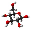
Jmol-3D images Image
Image
KEGG C00031 
MeSH Glucose
PubChem 5793
RTECS number LZ6600000
UNII 5SL0G7R0OK Yes
Properties
C6H12O6
Molar mass 180.16 g·mol−1
Appearance White powder
Density 1.54 g/cm3
Melting point α-D-glucose: 146 °C (295 °F; 419 K)
β-D-glucose: 150 °C (302 °F; 423 K)
909 g/1 L (25 °C (77 °F))
Thermochemistry
218.6 J K−1 mol−1[1]
209.2 J K−1 mol−1[1]
−1271 kJ/mol [2]
−2805 kJ/mol
Hazards
Safety data sheet ICSC 0865
NFPA 704
Flammability code 1: Must be pre-heated before ignition can occur. Flash point over 93 °C (200 °F). E.g., canola oil Health code 0: Exposure under fire conditions would offer no hazard beyond that of ordinary combustible material. E.g., sodium chloride Reactivity code 0: Normally stable, even under fire exposure conditions, and is not reactive with water. E.g., liquid nitrogen Special hazards (white): no code

NFPA 704 four-colored diamond

Except where otherwise noted, data are given for materials in their standard state (at 25 °C [77 °F], 100 kPa).
  verify (what isYes/?)
Infobox references

Function in biology[edit]

Glucose is the most widely used aldohexose in living organisms. One possible explanation for this is that glucose has a lower tendency than other aldohexoses to react non-specifically with the amine groups of proteins.[4] This reaction –glycation – impairs or destroys the function of many proteins.[4] Glucose’s low rate of glycation can be attributed to it having a more stable cyclic form compared to other aldohexoses, which means it spends less time than they do in its reactive open-chain form.[4] The reason for glucose having the most stable cyclic form of all the aldohexoses is due it having all of its hydroxy groups (with the exception of the hydroxy group on the anomeric carbon of D-glucose) in theequatorial position. Many of the long-term complications of diabetes (e.g., blindness, renal failure, and peripheral neuropathy) are probably due to the glycation of proteins or lipids.[5] In contrast, enzyme-regulated addition of sugars to protein is called glycosylation and is essential for the function of many proteins.[6]

Analyte in medical blood test[edit]

Main article: Glucose test

Glucose is a common medical analyte measured in blood samples. Eating or fasting prior to taking a blood sample has an effect on the result. A high fasting glucose blood sugar level may be a sign of prediabetes or diabetes mellitus.

Energy source[edit]

Glucose is a ubiquitous fuel in biology. It is used as an energy source in most organisms, from bacteria to humans, through either aerobic respiration, anaerobic respiration, or fermentation. Glucose is the human body’s key source of energy, through aerobic respiration, providing about 3.75 kilocalories (16 kilojoules) of food energy per gram.[7]Breakdown of carbohydrates (e.g. starch) yields mono- and disaccharides, most of which is glucose. Throughglycolysis and later in the reactions of the citric acid cycle and oxidative phosphorylation, glucose is oxidized to eventually form CO2 and water, yielding energy mostly in the form of ATP. The insulin reaction, and other mechanisms, regulate the concentration of glucose in the blood.

Glucose is a primary source of energy for the brain, so its availability influences psychological processes. Whenglucose is low, psychological processes requiring mental effort (e.g., self-control, effortful decision-making) are impaired.[8][9][10][11]

Glycolysis[edit]

α-D-Glucose Hexokinase α-DGlucose-6-phosphate
D-glucose wpmp.svg Alpha-D-glucose-6-phosphate wpmp.svg
ATP ADP
Biochem reaction arrow forward YYNN horiz med.png
Compound C00031 at KEGG Pathway Database. Enzyme 2.7.1.1 atKEGG Pathway Database. Compound C00668 at KEGG Pathway Database. Reaction R01786 at KEGG Pathway Database.

Glucose metabolism and various forms of it in the process
Glucose-containing compounds andisomeric forms are digested and taken up by the body in the intestines, including starch, glycogen,disaccharides and monosaccharides.
Glucose is stored in mainly the liver and muscles as glycogen.
It is distributed and used in tissues as free glucose.

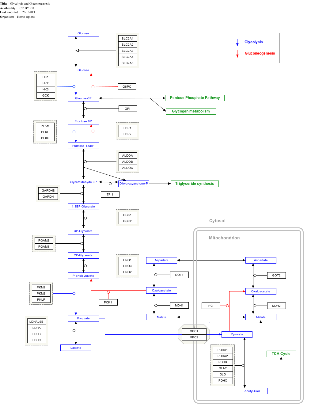
Use of glucose as an energy source in cells is by either aerobic respiration, anaerobic respiration, or fermentation. All of these processes follow from an earlier metabolic pathway known as glycolysis. The first step of glycolysis is thephosphorylation of glucose by a hexokinase to form glucose 6-phosphate. The main reason for the immediate phosphorylation of glucose is to prevent its diffusion out of the cell as the charged phosphate group prevents glucose 6-phosphate from easily crossing the cell membrane. Furthermore, addition of the high-energy phosphate groupactivates glucose for subsequent breakdown in later steps of glycolysis. At physiological conditions this initial reaction is irreversible.

In anaerobic respiration, one glucose molecule produces a net gain of two ATP molecules (four ATP molecules are produced during glycolysis, but two are required by enzymes used during the process).[12] In aerobic respiration, a molecule of glucose is much more profitable in that a maximum net production of 30 or 32 ATP molecules (depending on the organism) is generated.[13]

Click on genes, proteins and metabolites below to link to respective articles. [§ 1]

[[File:

GlycolysisGluconeogenesis_WP534

go to article go to article go to article go to article go to article go to article go to article go to article go to article go to article go to article go to article go to article go to article go to article go to article go to article go to article go to article go to article go to article go to article go to article go to article go to article go to article go to article go to article go to article go to article go to article go to article go to article go to article go to article go to article go to article go to article go to article go to article go to article go to article go to article go to article go to article go to article go to article go to article go to article go to article go to article go to article go to article go to article go to article go to article go to article go to article go to article go to article go to article go to article go to article go to article go to Entrez go to article go to article go to article go to article go to article go to WikiPathways go to article go to Entrez go to article
[[

]]

[[

]]

[[

]]

[[

]]

[[

]]

[[

]]

[[

]]

[[

]]

[[

]]

[[

]]

[[

]]

[[

]]

[[

]]

[[

]]

[[

]]

[[

]]

[[

]]

[[

]]

[[

]]

[[

]]

[[

]]

[[

]]

[[

]]

[[

]]

[[

]]

[[

]]

[[

]]

[[

]]

[[

]]

[[

]]

[[

]]

[[

]]

[[

]]

[[

]]

[[

]]

[[

]]

[[

]]

[[

]]

[[

]]

[[

]]

[[

]]

[[

]]

[[

]]

[[

]]

[[

]]

[[

]]

[[

]]

[[

]]

[[

]]

[[

]]

[[

]]

[[

]]

[[

]]

[[

]]

[[

]]

[[

]]

[[

]]

[[

]]

[[

]]

[[

]]

[[

]]

[[

]]

[[

]]

[[

]]

[[

]]

[[

]]

[[

]]

[[

]]

[[

]]

[[

]]

[[

]]

[[

]]

[[

]]

[[

]]

[[

]]

GlycolysisGluconeogenesis_WP534

go to article go to article go to article go to article go to article go to article go to article go to article go to article go to article go to article go to article go to article go to article go to article go to article go to article go to article go to article go to article go to article go to article go to article go to article go to article go to article go to article go to article go to article go to article go to article go to article go to article go to article go to article go to article go to article go to article go to article go to article go to article go to article go to article go to article go to article go to article go to article go to article go to article go to article go to article go to article go to article go to article go to article go to article go to article go to article go to article go to article go to article go to article go to article go to article go to Entrez go to article go to article go to article go to article go to article go to WikiPathways go to article go to Entrez go to article

|{{{bSize}}}px|alt=Glycolysis and Gluconeogenesis edit]]

Glycolysis and Gluconeogenesis edit

  1. Jump up^ The interactive pathway map can be edited at WikiPathways: “GlycolysisGluconeogenesis_WP534”.

Precursor[edit]

Organisms use glucose as a precursor for the synthesis of several important substances. Starch, cellulose, and glycogen (“animal starch”) are common glucosepolymers (polysaccharides). Some of these polymers (starch or glycogen) serve as energy stores, while others (cellulose and chitin, which is made from a derivative of glucose) have structural roles. Oligosaccharides of glucose combined with other sugars serve as important energy stores. These include lactose, the predominant sugar in milk, which is a glucose-galactose disaccharide, and sucrose, another disaccharide which is composed of glucose and fructose. Glucose is also added onto certain proteins and lipids in a process called glycosylation. This is often critical for their functioning. The enzymes that join glucose to other molecules usually use phosphorylated glucose to power the formation of the new bond by coupling it with the breaking of the glucose-phosphate bond.

Other than its direct use as a monomer, glucose can be broken down to synthesize a wide variety of other biomolecules. This is important, as glucose serves both as a primary store of energy and as a source of organic carbon. Glucose can be broken down and converted into lipids. It is also a precursor for the synthesis of other important molecules such as vitamin C (ascorbic acid).

Structure and nomenclature[edit]

Glucose is a monosaccharide with formula C6H12O6 or H-(C=O)-(CHOH)5-H, whose five hydroxyl (OH) groups are arranged in a specific way along its six-carbonback.

Open-chain form[edit]

Glucose can exist in both a straight-chain and ring form.

D-Glucose in Fischer projection

In its fleeting open-chain form, the glucose molecule has an open (as opposed to cyclic) and unbranched backbone of six carbon atoms, C-1 through C-6; where C-1 is part of an aldehyde group H(C=O)-, and each of the other five carbons bears one hydroxyl group -OH. The remaining bonds of the backbone carbons are satisfied by hydrogen atoms -H. Therefore glucose is both a hexose and an aldose, or analdohexose. The aldehyde group makes glucose a reducing sugar giving a positive reaction with theFehling test.

Each of the four carbons C-2 through C-5 is a stereocenter, meaning that its four bonds connect to four different substituents. (Carbon C-2, for example, connects to -(C=O)H, -OH, -H, and -(CHOH)4H.) In D-glucose, these four parts must be in a specific three-dimensional arrangement. Namely, when the molecule is drawn in the Fischer projection, the hydroxyls on C-2, C-4, and C-5 must be on the right side, while that on C-3 must be on the left side.

The positions of those four hydroxyls are exactly reversed in the Fischer diagram of L-glucose. D– and L-glucose are two of the 16 possible aldohexoses; the other 14 are allose, altrose, mannose, gulose, idose, galactose, and talose, each with two enantiomers, “D-” and “L-“.

Cyclic forms[edit]

In solutions, the open-chain form of glucose (either “D-” or “L-“) exists in equilibrium with several cyclic isomers, each containing a ring of carbons closed by one oxygen atom. In aqueous solution however, more than 99% of glucose molecules, at any given time, exist as pyranose. The open-chain form is limited to about 0.25% and furanose exists in negligible amounts. The terms “glucose” and “D-glucose” are generally used for these cyclic forms as well. The ring arises from the open-chain form by a nucleophilic addition reaction between the aldehyde group -(C=O)H at C-1 and the hydroxyl group -OH at C-4 or C-5, yielding a hemiacetalgroup -C(OH)H-O-.

The reaction between C-1 and C-5 creates a molecule with a six-membered ring, called pyranose, after the cyclic ether pyran, the simplest molecule with the same carbon-oxygen ring. The (much rarer) reaction between C-1 and C-4 creates a molecule with a five-membered ring, called furanose, after the cyclic ether furan. In either case, each carbon in the ring has one hydrogen and one hydroxyl attached, except for the last carbon (C-4 or C-5) where the hydroxyl is replaced by the remainder of the open molecule (which is -(C(CH2OH)HOH)-H or -(CHOH)-H, respectively).

The ring-closing reaction makes carbon C-1 chiral, too, since its four bonds lead to -H, to -OH, to carbon C-2, and to the ring oxygen. These four parts of the molecule may be arranged around C-1 (the anomeric carbon) in two distinct ways, designated by the prefixes “α-” and “β-“. When a glucopyranose molecule is drawn in the Haworth projection, the designation “α-” means that the hydroxyl group attached to C-1 and the -CH2OH group at C-5 lies on opposite sides of the ring’s plane (a trans arrangement), while “β-” means that they are on the same side of the plane (a cis arrangement).

Therefore, the open-chain isomer D-glucose gives rise to four distinct cyclic isomers: α-D-glucopyranose, β-D-glucopyranose, α-D-glucofuranose, and β-D-glucofuranose. These are all chiral.

Alpha-D-Glucopyranose.svgα-D
Glucopyranose
Beta-D-Glucopyranose.svgβ-D
Glucopyranose
Alpha-D-Glucofuranose.svgα-D
Glucofuranose
Beta-D-Glucofuranose.svgβ-D
Glucofuranose

The other open-chain isomer L-glucose similarly gives rise to four distinct cyclic forms of L-glucose, each the mirror image of the corresponding D-glucose.

The rings are not planar, but are twisted in three dimensions. The glucopyranose ring (α or β) can assume several non-planar shapes, analogous to the “chair” and “boat” conformations of cyclohexane. Similarly, the glucofuranose ring may assume several shapes, analogous to the “envelope” conformations of cyclopentane.

The glucopyranose forms of glucose predominate in solution, and are the only forms observed in the solid state. They are crystalline colorless solids, highly soluble in water and acetic acid, poorly soluble in methanol and ethanol. They melt at 146 °C (295 °F) (α) and 150 °C (302 °F) (β), and decompose at higher temperatures into carbon and water.

Rotational isomers[edit]

Each glucose isomer is subject to rotational isomerism. Within the cyclic form of glucose, rotation may occur around the O6-C6-C5-O5 torsion angle, termed the ω-angle, to form three staggered rotamer conformations called gauchegauche (gg), gauchetrans (gt) and transgauche (tg). For methyl α-D-glucopyranose at equilibrium the ratio of molecules in each rotamer conformation is reported as 57:38:5 gg:gt:tg.[14] This tendency for the ω-angle to prefer to adopt a gaucheconformation is attributed to the gauche effect.

Physical properties[edit]

Solutions[edit]

Glucose 5%

All forms of glucose are colorless and easily soluble in water, acetic acid, and several other solvents. They are only sparingly soluble in methanol and ethanol.

The open-chain form is thermodynamically unstable, and it spontaneously isomerizes to the cyclic forms. (Although the ring closure reaction could in theory create four- or three-atom rings, these would be highly strained, and are not observed in practice.) In solutions at room temperature, the four cyclic isomers interconvert over a time scale of hours, in a process called mutarotation.[15] Starting from any proportions, the mixture converges to a stable ratio of α:β 36:64. The ratio would be α:β 11:89 if it were not for the influence of the anomeric effect.[16] Mutarotation is considerably slower at temperatures close to 0 °C (32 °F).

Mutarotation consists of a temporary reversal of the ring-forming reaction, resulting in the open-chain form, followed by a reforming of the ring. The ring closure step may use a different -OH group than the one recreated by the opening step (thus switching between pyranose and furanose forms), and/or the new hemiacetal group created on C-1 may have the same or opposite handedness as the original one (thus switching between the α and β forms). Thus, though the open-chain form is barely detectable in solution, it is an essential component of the equilibrium.

Solid state[edit]

Depending on conditions, three major solid forms of glucose can be crystallised from water solutions: α-glucopyranose, β-glucopyranose, and β-glucopyranose hydrate.[17]

Optical activity[edit]

Whether in water or in the solid form, D-glucose is dextrorotatory, meaning it will rotate the direction of polarized light clockwise. The effect is due to the chirality of the molecules, and indeed the mirror-image isomer, L-glucose, is levorotatory (rotates polarized light counterclockwise) by the same amount. The strength of the effect is different for each of the five tautomers.

Note that the D– prefix does not refer directly to the optical properties of the compound. It indicates that the C-2 chiral center has the same handedness as that of D-glyceraldehyde (which was so labeled because it is dextrorotatory). The fact that D-glucose is dextrorotatory is a combined effect of its four chiral centers, not just of C-2; and indeed some of the other D-aldohexoses are levorotatory.

Computational Models[edit]

Glucose physical and chemical properties have been used to build a computational model of protein-glucose binding-sites.[18]

Production[edit]

Biosynthesis[edit]

In plants and some prokaryotes, glucose is a product of photosynthesis. In animals and fungi, glucose results from the breakdown of glycogen, a process known asglycogenolysis. In plants the breakdown substrate is starch.

In animals, glucose is synthesized in the liver and kidneys from non-carbohydrate intermediates, such as pyruvate, lactate and glycerol, by a process known asgluconeogenesis.

In some deep-sea bacteria, glucose is produced by chemosynthesis.

Commercial[edit]

Glucose is produced commercially via the enzymatic hydrolysis of starch. Many crops can be used as the source of starch. Maize, rice, wheat, cassava, corn huskand sago are all used in various parts of the world. In the United States, corn starch (from maize) is used almost exclusively. Most commercial glucose occurs as a component of invert sugar, a roughly 1:1 mixture of glucose and fructose. In principle, cellulose could be hydrolysed to glucose, but this process is not yet commercially practical.[17] Glucose has approximately 75% the sweetness of sucrose (table sugar).[19]

Sources and absorption[edit]

Most dietary carbohydrates contain glucose, either as their only building block, as in starch and glycogen, or together with another monosaccharide, as in sucrose and lactose.

In the lumen of the duodenum and small intestine, the glucose oligo- and polysaccharides are broken down to monosaccharides by the pancreatic and intestinal glycosidases. Other polysaccharides cannot be processed by the human intestine and require assistance by intestinal flora if they are to be broken down; the most notable exceptions are sucrose (fructose-glucose) and lactose (galactose-glucose). Glucose is then transported across the apical membrane of the enterocytes bySLC5A1 (SGLT1), and later across their basal membrane by SLC2A2 (GLUT2).[20] Some of the glucose is converted to lactic acid by astrocytes, which is then utilized as an energy source by brain cells; some of the glucose is used by intestinal cells and red blood cells, while the rest reaches the liver, adipose tissue andmuscle cells, where it is absorbed and stored as glycogen (under the influence of insulin). Liver cell glycogen can be converted to glucose and returned to the blood when insulin is low or absent; muscle cell glycogen is not returned to the blood because of a lack of enzymes. In fat cells, glucose is used to power reactions that synthesize some fat types and have other purposes. Glycogen is the body’s “glucose energy storage” mechanism, because it is much more “space efficient” and less reactive than glucose itself.

Glucose tablets

In hypoglycemia management[edit]

Individuals with diabetes or other conditions where hypoglycemia (low blood sugar) may occur often carry small amounts of sugar in various forms. One sugar commonly used is glucose, often in the form of glucose tablets (glucose pressed into a tablet shape sometimes with one or more other ingredients as a binder).

History[edit]

Glucose was first isolated (from raisins) in 1747 by the German chemist Andreas Marggraf.[21] Because glucose is a basic necessity of many organisms, a correct understanding of its chemical makeup and structure contributed greatly to a general advancement in organic chemistry. This understanding occurred largely as a result of the investigations of Emil Fischer, a German chemist who received the 1902 Nobel Prize in Chemistry for his findings.[22] The synthesis of glucose established the structure of organic material and consequently formed the first definitive validation of Jacobus Henricus van’t Hoff‘s theories of chemical kinetics and the arrangements of chemical bonds in carbon-bearing molecules.[23] Between 1891 and 1894, Fischer established the stereochemical configuration of all the known sugars and correctly predicted the possibleisomers, applying van’t Hoff’s theory of asymmetrical carbon atoms.

How to add fractions

There are 3 Simple Steps to add fractions:

  • Step 1: Make sure the bottom numbers (the denominators) are the same
  • Step 2: Add the top numbers (the numerators), put the answer over thedenominator
  • Step 3: Simplify the fraction (if needed)

Example 1:

1  +  1
4 4

Step 1. The bottom numbers (the denominators) are already the same. Go straight to step 2.

Step 2. Add the top numbers and put the answer over the same denominator:

1  +  1  =  1 + 1  =  2
4 4 4 4

Step 3. Simplify the fraction:

2  =  1
4 2

In picture form it looks like this:

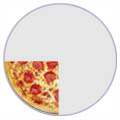
1/4 + 1/4 = 2/4 = 1/2
1/4 1/4 2/4 1/2

… and do you see how 2/4 is simpler as 1/2 ? (see Equivalent Fractions.)

Example 2:

1  +  1
3 6

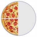
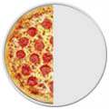
Step 1: The bottom numbers are different. See how the slices are different sizes?

1/3 + 1/6 = ?
1/3 1/6 1 1

We need to make them the same before we can continue, because we can’t add them like that.

The number “6” is twice as big as “3”, so to make the bottom numbers the same we can multiply the top and bottom of the first fraction by 2, like this:

× 2
1   =   2
3 6
× 2

Important: you multiply both top and bottom by the same amount,
to keep the value of the fraction the same

Now the fractions have the same bottom number (“6”), and our question looks like this:

2/6 + 1/6
2/6 1/6 1 1

The bottom numbers are now the same, so we can go to step 2.

Step 2: Add the top numbers and put them over the same denominator:

2  + 1 = 2 + 1 = 3
6 6 6 6

In picture form it looks like this:

2/6 + 1/6 = 3/6
2/6 1/6 3/6 1

Step 3: Simplify the fraction:

3  = 1
6 2

In picture form the whole answer looks like this:

2/6 + 1/6 = 3/6 = 1/2
2/6 1/6 3/6 1/2

With Pen and Paper

And here is how to do it with a pen and paper (press the play button):

Play with it!

Try the Adding Fractions Animation.

A Rhyme To Help You Remember

“If adding or subtracting is your aim,
The bottom numbers must be the same!

“Change the bottom using multiply or divide,
But the same to the top must be applied,

“And don’t forget to simplify,
Before its time to say good bye”

Example 3:

1  +  1
3 5

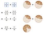
Again, the bottom numbers are different (the slices are different sizes)!

1/3 + 1/5 = ?
1/3 1/5 1 1

But let us try dividing them into smaller sizes that will each be the same:

5/15 + 3/15
5/15 3/15 1 1

The first fraction: by multiplying the top and bottom by 5 we ended up with 5/15 :

× 5
1   =   5
3 15
× 5

The second fraction: by multiplying the top and bottom by 3 we ended up with 3/15 :

× 3
1   =   3
5 15
× 3

The bottom numbers are now the same, so we can go ahead and add the top numbers:

5/15 + 3/15 = 8/15
5/15 3/15 8/15 1

The result is already as simple as it can be, so that is the answer: 8/15

Making the Denominators the Same

In the previous example how did we know to cut them into 1/15ths to make the denominators the same? Read how to do this using either one of these methods:

They both work, use which one you prefer!

Example: Cupcakes

You want to make and sell cupcakes:

  • A friend can supply the ingredients, if you give them1/3 of sales
  • And a market stall costs 1/4 of sales

How much is that altogether?

We need to add 1/3 and 1/4

1 + 1 = ?
3 4 ?

First make the bottom numbers (the denominators) the same.

Multiply top and bottom of 1/3 by 4:

1 × 4 + 1 = ?
3 × 4 4 ?

And multiply top and bottom of 1/4 by 3:

1 × 4 + 1 × 3 = ?
3 × 4 4 × 3 ?

Now do the calculations:

4 + 3 = 4+3 = 7
12 12 12 12

Answer: 7/12 of sales go in ingredients and market costs.

Why is the sky blue?

Why is the sky blue?

A clear cloudless day-time sky is blue because molecules in the air scatter blue light from the sun more than they scatter red light.  When we look towards the sun at sunset, we see red and orange colours because the blue light has been scattered out and away from the line of sight.

The white light from the sun is a mixture of all colours of the rainbow.  This was demonstrated by Isaac Newton, who used a prism to separate the different colours and so form a spectrum.  The colours of light are distinguished by their different wavelengths.  The visible part of the spectrum ranges from red light with a wavelength of about 720 nm, to violet with a wavelength of about 380 nm, with orange, yellow, green, blue and indigo between.  The three different types of colour receptors in the retina of the human eye respond most strongly to red, green and blue wavelengths, giving us our colour vision.

Tyndall Effect

The first steps towards correctly explaining the colour of the sky were taken by John Tyndall in 1859.  He discovered that when light passes through a clear fluid holding small particles in suspension, the shorter blue wavelengths are scattered more strongly than the red.  This can be demonstrated by shining a beam of white light through a tank of water with a little milk or soap mixed in.  From the side, the beam can be seen by the blue light it scatters; but the light seen directly from the end is reddened after it has passed through the tank.  The scattered light can also be shown to be polarised using a filter of polarised light, just as the sky appears a deeper blue through polaroid sun glasses.

This is most correctly called the Tyndall effect, but it is more commonly known to physicists as Rayleigh scattering—after Lord Rayleigh, who studied it in more detail a few years later.  He showed that the amount of light scattered is inversely proportional to the fourth power of wavelength for sufficiently small particles.  It follows that blue light is scattered more than red light by a factor of (700/400)4 ~= 10.

Dust or Molecules?

Tyndall and Rayleigh thought that the blue colour of the sky must be due to small particles of dust and droplets of water vapour in the atmosphere.  Even today, people sometimes incorrectly say that this is the case.  Later scientists realised that if this were true, there would be more variation of sky colour with humidity or haze conditions than was actually observed, so they supposed correctly that the molecules of oxygen and nitrogen in the air are sufficient to account for the scattering.  The case was finally settled by Einstein in 1911, who calculated the detailed formula for the scattering of light from molecules; and this was found to be in agreement with experiment.  He was even able to use the calculation as a further verification of Avogadro’s number when compared with observation.  The molecules are able to scatter light because the electromagnetic field of the light waves induces electric dipole moments in the molecules.

Why not violet?

If shorter wavelengths are scattered most strongly, then there is a puzzle as to why the sky does not appear violet, the colour with the shortest visible wavelength.  The spectrum of light emission from the sun is not constant at all wavelengths, and additionally is absorbed by the high atmosphere, so there is less violet in the light.  Our eyes are also less sensitive to violet.  That’s part of the answer; yet a rainbow shows that there remains a significant amount of visible light coloured indigo and violet beyond the blue.  The rest of the answer to this puzzle lies in the way our vision works.  We have three types of colour receptors, or cones, in our retina.  They are called red, blue and green because they respond most strongly to light at those wavelengths.  As they are stimulated in different proportions, our visual system constructs the colours we see.


Response curves for the three types of cone in the human eye

When we look up at the sky, the red cones respond to the small amount of scattered red light, but also less strongly to orange and yellow wavelengths.  The green cones respond to yellow and the more strongly scattered green and green-blue wavelengths.  The blue cones are stimulated by colours near blue wavelengths, which are very strongly scattered.  If there were no indigo and violet in the spectrum, the sky would appear blue with a slight green tinge.  However, the most strongly scattered indigo and violet wavelengths stimulate the red cones slightly as well as the blue, which is why these colours appear blue with an added red tinge.  The net effect is that the red and green cones are stimulated about equally by the light from the sky, while the blue is stimulated more strongly.  This combination accounts for the pale sky blue colour.  It may not be a coincidence that our vision is adjusted to see the sky as a pure hue.  We have evolved to fit in with our environment; and the ability to separate natural colours most clearly is probably a survival advantage.


A multicoloured sunset over the Firth of Forth in Scotland.

Sunsets

When the air is clear the sunset will appear yellow, because the light from the sun has passed a long distance through air and some of the blue light has been scattered away.  If the air is polluted with small particles, natural or otherwise, the sunset will be more red.  Sunsets over the sea may also be orange, due to salt particles in the air, which are effective Tyndall scatterers.  The sky around the sun is seen reddened, as well as the light coming directly from the sun.  This is because all light is scattered relatively well through small angles—but blue light is then more likely to be scattered twice or more over the greater distances, leaving the yellow, red and orange colours.


A blue haze over the mountains of Les Vosges in France.

Blue Haze and Blue Moon

Clouds and dust haze appear white because they consist of particles larger than the wavelengths of light, which scatter all wavelengths equally (Mie scattering).  But sometimes there might be other particles in the air that are much smaller.  Some mountainous regions are famous for their blue haze.  Aerosols of terpenes from the vegetation react with ozone in the atmosphere to form small particles about 200 nm across, and these particles scatter the blue light.  A forest fire or volcanic eruption may occasionally fill the atmosphere with fine particles of 500—800 nm across, being the right size to scatter red light.  This gives the opposite to the usual Tyndall effect, and may cause the moon to have a blue tinge since the red light has been scattered out.  This is a very rare phenomenon, occurring literally once in a blue moon.

Opalescence

The Tyndall effect is responsible for some other blue coloration’s in nature: such as blue eyes, the opalescence of some gem stones, and the colour in the blue jay’s wing.  The colours can vary according to the size of the scattering particles.  When a fluid is near its critical temperature and pressure, tiny density fluctuations are responsible for a blue coloration known as critical opalescence.  People have also copied these natural effects by making ornamental glasses impregnated with particles, to give the glass a blue sheen.  But not all blue colouring in nature is caused by scattering.  Light under the sea is blue because water absorbs longer wavelength of light through distances over about 20 metres.  When viewed from the beach, the sea is also blue because it reflects the sky, of course.  Some birds and butterflies get their blue colorations by diffraction effects.

Why is the Mars sky red?

Images sent back from the Viking Mars landers in 1977 and from Pathfinder in 1997 showed a red sky seen from the Martian surface.  This was due to red iron-rich dusts thrown up in the dust storms occurring from time to time on Mars.  The colour of the Mars sky will change according to weather conditions.  It should be blue when there have been no recent storms, but it will be darker than the earth’s daytime sky because of Mars’ thinner atmosphere.

Stingray

Stingrays are a group of rays, which are cartilaginous fish related to sharks. They are classified in the suborderMyliobatoidei of the order Myliobatiformes and consist of eight families: Hexatrygonidae (sixgill stingray), Plesiobatidae(deep water stingray), Urolophidae (stingarees), Urotrygonidae (round rays), Dasyatidae (whiptail stingrays),Potamotrygonidae (river stingrays), Gymnuridae (butterfly rays), and Myliobatidae (eagle rays).[1][2]

Most stingrays have one or more barbed stingers (modified from dermal denticles) on the tail, which are used exclusively in self-defense. The stinger may reach a length of approximately 35 cm (14 in), and its underside has two grooves with venomglands.[3] The stinger is covered with a thin layer of skin, the integumentary sheath, in which the venom is concentrated.[4] A few members of the suborder, such as the manta and porcupine rays, do not have stingers.[5]

Stingrays are common in coastal tropical and subtropical marine waters throughout the world. Some species, such asDasyatis thetidis, are found in warmer temperate oceans, and others, such as Plesiobatis daviesi, are found in the deep ocean. The river stingrays, and a number of whiptail stingrays (such as the Niger stingray), are restricted to fresh water. Most myliobatoids are demersal (inhabiting the next-to-lowest zone in the water column); but some, such as the pelagic stingray and the eagle rays, are pelagic.[6]

While most stingrays are relatively widespread and not currently threatened, for several species (for example Taeniura meyeni, D. colarensis, D. garouaensis, and D. laosensis), conservation status is more problematic, leading to their being listed as vulnerable or endangered by IUCN. The status of several other species are poorly known, leading to their being listed as Data Deficient.[7]

Stingrays
Temporal range: Early Cretaceous–Recent[1]

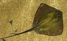
Dasyatis pastinaca01.jpg
Common stingray (Dasyatis pastinaca)
Scientific classification
Kingdom: Animalia
Phylum: Chordata
Class: Chondrichthyes
Subclass: Elasmobranchii
Order: Myliobatiformes
Suborder: Myliobatoidei
Families

Behavior[edit]

A stingray’s underside shows its mouth and the ventral gill slits. The pair of claspers (at the base of the tail) identifies this individual as male.

The flattened bodies of stingrays allow them to effectively conceal themselves in their environment. Stingrays do this by agitating the sand and hiding beneath it. Because their eyes are on top of their bodies and their mouths on the undersides, stingrays cannot see their prey; instead, they use smell and electroreceptors (ampullae of Lorenzini) similar to those ofsharks.[8] Stingrays feed primarily on molluscs, crustaceans, and occasionally on small fish. Some stingrays’ mouths contain two powerful, shell-crushing plates, while other species only have sucking mouthparts. Stingrays settle on the bottom while feeding, often leaving only their eyes and tail visible. Coral reefs are favorite feeding grounds and are usually shared with sharks during high tide.[9]

Reproduction[edit]

When a male is courting a female, he will follow her closely, biting at her pectoral disc. He then places one of his two claspers into her valve.[10]

Stingrays are ovoviviparous, bearing live young in “litters” of 5 to 13. The female holds the embryos in the womb without a placenta. Instead, the embryos absorb nutrients from a yolk sac, and after the sac is depleted, the mother provides uterine “milk”.[11]

At the Sea Life London Aquarium, two female stingrays have delivered seven baby stingrays, although the mothers have not been near a male for two years. “Rays have been known to store sperm and not give birth until they decide the timing is right”.[12]

Diet[edit]

A stingray’s diet includes small fish, snails, clams, and shrimp, and some other small sea creatures.[13]

Stingray injuries[edit]

Main article: Stingray injury

A stingray’s stinger (ruler in cm)

Stingrays do not aggressively attack humans, though stings do normally occur if a ray is accidentally stepped on.[14] To avoid stepping on a stingray in shallow water, the water should be waded through with a shuffle.[15] Alternatively, before wading, stones can be thrown into the water to scare stingrays away.[16] Contact with the stinger causes local trauma (from the cut itself), pain, swelling, muscle cramps from the venom, and later may result in infection from bacteria or fungus.[17] The injury is very painful, but seldom life-threatening unless the stinger pierces a vital area.[14] The barb usually breaks off in the wound, and surgery may be required to remove the fragments.[18]

Fatal stings are very rare, but can happen,[14] most famously in the death of Steve Irwin in 2006, in which the stinger penetrated the thoracic wall, causing massive trauma.[19]

As food[edit]

Rays are edible, and may be caught as food using fishing lines or spears.[20] Stingray recipes abound throughout the world, with dried forms of the wings being most common. For example, in Malaysia and Singapore, stingray is commonly grilled over charcoal, then served with spicy sambal sauce, or soysauce. Generally, the most prized parts of the stingray are the wings (flaps is the proper terminology), the “cheek” (the area surrounding the eyes), and the liver. The rest of the ray is considered too rubbery to have any culinary uses.[21]

Stingray teeth and jaws, on display at the American Museum of Natural History.

While not independently valuable as a food source, the stingray’s capacity to damage shell fishing grounds can lead to bounties being placed on their removal.[22]

Ecotourism[edit]

Stingrays are usually very docile and curious, their usual reaction being to flee any disturbance, but they will sometimes brush their fins past any new object they encounter. Nevertheless, certain larger species may be more aggressive and should be approached with caution, as the stingray’s defensive reflex (use of its poisoned stinger) may result in serious injury or death.[23]

Dasyatids are not normally visible to swimmers, but divers and snorkelers may find them in shallow, sandy waters, more so when the water is warm. In the Cayman Islands several dive sites called Stingray City, Grand Cayman, allow divers and snorkelers to swim with large southern stingrays (D. americana) and feed them by hand.[24] A “Stingray City” in the sea surrounding the Caribbean island of Antigua consists of a large, shallow reserve where the rays live, and snorkeling is possible, since the rays are used to the presence of humans.[25]

In Belize, off the island of Ambergris Caye, there is a popular marine sanctuary, Hol Chan, where divers and snorkelers often gather to watch stingrays and nurse sharks drawn to the area by tour operators who feed the animals.

Many Tahitian island resorts regularly offer guests the chance to “feed the stingrays and sharks”. This consists of taking a boat to the outer lagoon reefs, then standing in waist-high water while habituated stingrays swarm around, pressing right up against tourists seeking food from their hands or that being tossed into the water. The boat owners also “call in” sharks, which, when they arrive from the ocean, swoop through the shallow water above the reef and snatch food offered to them.[26]

Other uses[edit]

The skin of the ray (same in Japanese) is used as an under layer for the cord or leather wrap (known as ito in Japanese) on Japanese swords due to its hard, rough, skin texture that keeps the braided wrap from sliding on the handle during use. They are also used to make exotic shoes, boots, belts, wallets, jackets, and cellphone cases.[27]

Several ethnological sections in museums,[28] such as the British Museum, display arrowheads and spearheads made of stingray stingers, used in Micronesia and elsewhere.[29] Henry de Monfreid stated in his books that before World War II, in the Horn of Africa, whips were made from the tail of big stingrays, and these devices inflicted cruel cuts, so in Aden the British forbade their use on women and slaves. In former Spanish colonies, a stingray is called raya látigo (“whip ray”).

Monfreid also wrote in several places about men of his crew suffering stingray wounds while standing and wading into Red Sea shallows to load or unload smuggled wares: he wrote that to “save the man’s life”, searing the wound with a red-hot iron was necessary.[30]

Fossils[edit]

Although stingray teeth are rare on sea bottoms compared to the similar shark teeth, scuba divers searching for the latter do encounter the teeth of stingrays.Permineralized stingray teeth have been found in sedimentary deposits around the world, including fossiliferous outcrops in Morocco.[31]

Seth Rollins vs. Sting Announced for WWE Night of Champions 2015 PPV

The wait for Sting’s second match in a WWE ring may not be anywhere near as long as the wait for his first. The six-time WCW world champion returned during Monday night’s episode of Raw.

Triple H and Stephanie McMahon were planning to unveil a statue of Seth Rollins after he defeated John Cena at SummerSlam on Sunday night. Instead, The Icon was waiting behind the curtain, per the WWE:

After fans speculated for years about whether he’d ever make the jump to WWE, Sting shocked the wrestling world when he made his first appearance with the company during Survivor Series 2014. That kicked off a feud with Triple H that culminated in defeat at WrestleMania 31.

After the Raw immediately following WrestleMania, Sting gave a somewhat vague interview about what the future held for him in the company:

n the days leading up to WrestleMania 31, Sting didn’t shut the door completely on another WWE run, but he signaled his match with Triple H could be more of a one-off occurrence.

“I’m feeling like this is probably going to be my last one,” he said, perDan Gelston of the Associated Press. “We’ll see how everyone reacts. If I’m asked to do one more at Survivor Series or maybe even WrestleMania next year, at this point, I would consider it. But realistically, this is going to be my last one.”

Given Sting’s status in the industry, his list of potential challengers was pretty small. He’s not like Chris Jericho, who can randomly turn up at house shows and wrestle Neville on a WWE Network exclusive.

Whatever WWE had planned for Sting, it was going to be big.

It is a bit surprising the company isn’t teasing a match between Sting and The Undertaker. That was a natural pairing after years of fantasy booking by wrestling fans.

But Rollins is a strong replacement and an in-ring foil befitting Sting’s legendary status in the business. Plus, it continues the Sting vs. The Authority story arc that began at Survivor Series.

Kevin Sullivan, author of WWE 50, also envisions Sting elevating Rollins:

Nobody will expect Sting to be able to put on a five-star mat classic at 56 years old. His WrestleMania clash with Triple H wasn’t all that memorable beyond the interference from D-Generation X and the nWo.

That shouldn’t preclude Sting and Rollins from putting on a good match, however. Seeing Sting in a WWE ring for what could be the final time will be more than enough to make their fight a massive spectacle.

Proton

The proton is a subatomic particle, symbol p or p+, with a positive electric charge of +1e elementary charge and mass slightly less than that of a neutron. Protons and neutrons, each with mass approximately one atomic mass unit, are collectively referred to as “nucleons”. One or more protons are present in the nucleus of an atom. The number of protons in the nucleus is referred to as its atomic number. Since each element has a unique number of protons, each element has its own unique atomic number. The word proton is Greek for “first”, and this name was given to the hydrogen nucleus by Ernest Rutherford in 1920. In previous years Rutherford had discovered that the hydrogen nucleus (known to be the lightest nucleus) could be extracted from the nuclei of nitrogen by collision. The proton was therefore a candidate to be a fundamental particle and a building block of nitrogen and all other heavier atomic nuclei.
In the modern Standard Model of particle physics, the proton is a hadron, and like the neutron, the other nucleon (particle present in atomic nuclei), is composed of three quarks. Although the proton was originally considered a fundamental particle, it is composed of three valence quarks: two up quarks and one down quark. The rest masses of the quarks contribute only about 1% of the proton’s mass, however.[2] The remainder of the proton mass is due to the kinetic energy of the quarks and to the energy of the gluon fields that bind the quarks together. Because the proton is not a fundamental particle, it possesses a physical size; the radius of the proton is about 0.84–0.87 fm.[3]

At sufficiently low temperatures, free protons will bind to electrons. However, the character of such bound protons does not change, and they remain protons. A fast proton moving through matter will slow by interactions with electrons and nuclei, until it is captured by the electron cloud of an atom. The result is a protonated atom, which is a chemical compound of hydrogen. In vacuum, when free electrons are present, a sufficiently slow proton may pick up a single free electron, becoming a neutral hydrogen atom, which is chemically a free radical. Such “free hydrogen atoms” tend to react chemically with many other types of atoms at sufficiently low energies. When free hydrogen atoms react with each other, they form neutral hydrogen molecules (H2), which are the most common molecular component of molecular clouds in interstellar space. Such molecules of hydrogen on Earth may then serve (among many other uses) as a convenient source of protons for accelerators (as used in proton therapy) and other hadron particle physics experiments that require protons to accelerate, with the most powerful and noted example being the Large Hadron Collider.

History

The concept of a hydrogen-like particle as a constituent of other atoms was developed over a long period. As early as 1815, William Prout proposed that all atoms are composed of hydrogen atoms (which he called “protyles”), based on a simplistic interpretation of early values of atomic weights (see Prout’s hypothesis), which was disproved when more accurate values were measured.[7]:39–42

Ernest Rutherford at the first Solvay Conference, 1911

In 1886, Eugen Goldstein discovered canal rays (also known as anode rays) and showed that they were positively charged particles (ions) produced from gases. However, since particles from different gases had different values of charge-to-mass ratio (e/m), they could not be identified with a single particle, unlike the negative electrons discovered by J. J. Thomson.
Following the discovery of the atomic nucleus by Ernest Rutherford in 1911, Antonius van den Broek proposed that the place of each element in the periodic table (its atomic number) is equal to its nuclear charge. This was confirmed experimentally by Henry Moseley in 1913 using X-ray spectra.
In 1917, (in experiments reported in 1919) Rutherford proved that the hydrogen nucleus is present in other nuclei, a result usually described as the discovery of the proton.[8] Rutherford had earlier learned to produce hydrogen nuclei as a type of radiation produced as a product of the impact of alpha particles on nitrogen gas, and recognize them by their unique penetration signature in air and their appearance in scintillation detectors. These experiments were begun when Rutherford had noticed that, when alpha particles were shot into air (mostly nitrogen), his scintillation detectors showed the signatures of typical hydrogen nuclei as a product. After experimentation Rutherford traced the reaction to the nitrogen in air, and found that when alphas were produced into pure nitrogen gas, the effect was larger. Rutherford determined that this hydrogen could have come only from the nitrogen, and therefore nitrogen must contain hydrogen nuclei. One hydrogen nucleus was being knocked off by the impact of the alpha particle, producing oxygen-17 in the process. This was the first reported nuclear reaction, 14N + α → 17O + p. (This reaction would later be observed happening directly in a cloud chamber in 1925).
Rutherford knew hydrogen to be the simplest and lightest element and was influenced by Prout’s hypothesis that hydrogen was the building block of all elements. Discovery that the hydrogen nucleus is present in all other nuclei as an elementary particle, led Rutherford to give the hydrogen nucleus a special name as a particle, since he suspected that hydrogen, the lightest element, contained only one of these particles. He named this new fundamental building block of the nucleus the proton, after the neuter singular of the Greek word for “first”, πρῶτον. However, Rutherford also had in mind the word protyle as used by Prout. Rutherford spoke at the British Association for the Advancement of Science at its Cardiff meeting beginning 24 August 1920.[9] Rutherford was asked by Oliver Lodge for a new name for the positive hydrogen nucleus to avoid confusion with the neutral hydrogen atom. He initially suggested both proton and prouton (after Prout).[10] Rutherford later reported that the meeting had accepted his suggestion that the hydrogen nucleus be named the “proton”, following Prout’s word “protyle”.[11] The first use of the word “proton” in the scientific literature appeared in 1920.[12]

In quantum chromodynamics, the modern theory of the nuclear force, most of the mass of the proton and the neutron is explained by special relativity. The mass of the proton is about 80–100 times greater than the sum of the rest masses of the quarks that make it up, while the gluons have zero rest mass. The extra energy of the quarks and gluons in a region within a proton, as compared to the rest energy of the quarks alone in the QCD vacuum, accounts for almost 99% of the mass. The rest mass of the proton is, thus, the invariant mass of the system of moving quarks and gluons that make up the particle, and, in such systems, even the energy of massless particles is still measured as part of the rest mass of the system.
Two terms are used in referring to the mass of the quarks that make up protons: current quark mass refers to the mass of a quark by itself, while constituent quark mass refers to the current quark mass plus the mass of the gluon particle field surrounding the quark.[18]:285–286 [19]:150–151 These masses typically have very different values. As noted, most of a proton’s mass comes from the gluons that bind the current quarks together, rather than from the quarks themselves. While gluons are inherently massless, they possess energy—to be more specific, quantum chromodynamics binding energy (QCBE)—and it is this that contributes so greatly to the overall mass of the proton (see mass in special relativity). A proton has a mass of approximately 938 MeV/c2, of which the rest mass of its three valence quarks contributes only about 9.4 MeV/c2; much of the remainder can be attributed to the gluons’ QCBE.[20][21][22]
The internal dynamics of the proton are complicated, because they are determined by the quarks’ exchanging gluons, and interacting with various vacuum condensates. Lattice QCD provides a way of calculating the mass of the proton directly from the theory to any accuracy, in principle. The most recent calculations[23][24] claim that the mass is determined to better than 4% accuracy, even to 1% accuracy (see Figure S5 in Dürr et al.[24]). These claims are still controversial, because the calculations cannot yet be done with quarks as light as they are in the real world. This means that the predictions are found by a process of extrapolation, which can introduce systematic errors.[25] It is hard to tell whether these errors are controlled properly, because the quantities that are compared to experiment are the masses of the hadrons, which are known in advance.
These recent calculations are performed by massive supercomputers, and, as noted by Boffi and Pasquini: “a detailed description of the nucleon structure is still missing because … long-distance behavior requires a nonperturbative and/or numerical treatment…”[26] More conceptual approaches to the structure of the proton are: the topological soliton approach originally due to Tony Skyrme and the more accurate AdS/QCD approach that extends it to include a string theory of gluons,[27] various QCD-inspired models like the bag model and the constituent quark model, which were popular in the 1980s, and the SVZ sum rules, which allow for rough approximate mass calculations.[28] These methods do not have the same accuracy as the more brute-force lattice QCD methods, at least not yet.

Atomic number Edit

In chemistry, the number of protons in the nucleus of an atom is known as the atomic number, which determines the chemical element to which the atom belongs. For example, the atomic number of chlorine is 17; this means that each chlorine atom has 17 protons and that all atoms with 17 protons are chlorine atoms. The chemical properties of each atom are determined by the number of (negatively charged) electrons, which for neutral atoms is equal to the number of (positive) protons so that the total charge is zero. For example, a neutral chlorine atom has 17 protons and 17 electrons, whereas a Cl− anion has 17 protons and 18 electrons for a total charge of −1.
All atoms of a given element are not necessarily identical, however, as the number of neutrons may vary to form different isotopes, and energy levels may differ forming different nuclear isomers. For example, there are two stable isotopes of chlorine: 35

17Cl with 35 − 17 = 18 neutrons and 37

17Cl with 37 − 17 = 20 neutrons.
Hydrogen ion Edit

See also: Hydron (chemistry)
Protium, the most common isotope of hydrogen, consists of one proton and one electron (it has no neutrons). The term “hydrogen ion” (H+

) implies that that H-atom has lost its one electron, causing only a proton to remain. Thus, in chemistry, the terms “proton” and “hydrogen ion” (for the protium isotope) are used synonymously

In chemistry, the term proton refers to the hydrogen ion, H+

. Since the atomic number of hydrogen is 1, a hydrogen ion has no electrons and corresponds to a bare nucleus, consisting of a proton (and 0 neutrons for the most abundant isotope protium 1

1H). The proton is a “bare charge” with only about 1/64,000 of the radius of a hydrogen atom, and so is extremely reactive chemically. The free proton, thus, has an extremely short lifetime in chemical systems such as liquids and it reacts immediately with the electron cloud of any available molecule. In aqueous solution, it forms the hydronium ion, H3O+, which in turn is further solvated by water molecules in clusters such as [H5O2]+ and [H9O4]+.[33]
The transfer of H+

 in an acid–base reaction is usually referred to as “proton transfer”. The acid is referred to as a proton donor and the base as a proton acceptor. Likewise, biochemical terms such as proton pump and proton channel refer to the movement of hydrated H+

 ions.
The ion produced by removing the electron from a deuterium atom is known as a deuteron, not a proton. Likewise, removing an electron from a tritium atom produces a triton.
Proton nuclear magnetic resonance (NMR) Edit

Also in chemistry, the term “proton NMR” refers to the observation of hydrogen-1 nuclei in (mostly organic) molecules by nuclear magnetic resonance. This method uses the spin of the proton, which has the value one-half. The name refers to examination of protons as they occur in protium (hydrogen-1 atoms) in compounds, and does not imply that free protons exist in the compound being studied.